The following white paper was written by our founder and medical director, Dr. Kenneth Harper, to share with physicians in Macon, Warner Robins, and the surrounding area. It is designed to help physicians improve speed and access to care in the event of an SVT.
If you are a physician, continue reading below.
If you are a patient, you are welcome to continue reading below. You may also contact us for an evaluation for your varicose veins, spider veins, and leg swelling, or you can visit our website to learn more about our practice and how we can help you. However, this article is not a substitute for you seeking emergency care if you are concerned that you might have a blood clot. In particular, if you are experiencing symptoms such as shortness of breath, tightness or heaviness in the chest, or a sense of impending doom, call 911 or go to your nearest emergency room. These are symptoms of a potentially life-threatening condition known as Pulmonary Embolism (PE).
 DVT Awareness Month: A New Era In The Diagnosis And Treatment Of Superficial Vein Thrombosis (SVT)
by: Kenneth Harper, MD, FACS, RPVI, RPhS., Vein Specialists of the South, LLC
A New Look At SVT
During Blood Clot Awareness Month, I want to share an update on Superficial Vein Thrombosis (SVT) and Deep Vein Thrombosis (DVT). As a bonus, I summarized the findings of a study linking varicose veins with an increased risk of venous thromboembolism (SVT/DVT/PE) and Peripheral Artery Disease (PAD). I also includes some information on DVT.
Why Worry If It Is Just An SVT?
For years, SVT was considered a self-limiting condition with few risks. In most cases, it was diagnosed with a clinical history and physical exam. However, new information now associates SVT with an increased risk of DVT and PE. The data has resulted in updated recommendations for the diagnosis and treatment of SVT.
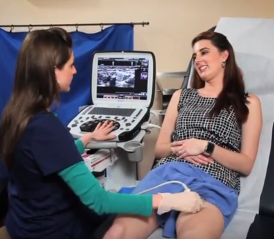
The bottom line: If you suspect a blood clot (SVT or DVT), a duplex ultrasound should be performed.
Virchow’s Triad And Blood Clots
Doctor Virchow identified three factors that increase the risk of developing a VTE. Virchow’s Triad includes:
- stagnant venous blood flow
- a hypercoagulable state which makes the blood more likely to clot
- an injury to the vein wall
Deep vein and superficial vein disease including varicose veins trigger Virchow’s Triad by slowing the venous blood return to the heart.
Venous Thrombo-Embolism (VTE) Complex
VTE includes SVT, DVT and PE:

- An SVT is a blood clot in the superficial veins within the arm, leg or trunk. CT scan data of patients with SVT has shown that up to one third of these patients have asymptomatic micro-pulmonary emboli.
- A DVT is a blood clot in the deep veins.
- A PE is when a clot breaks loose and travels to the lungs. The most likely source of clots causing a PE are from the deep veins in the legs and pelvis. A PE can be life threatening. Someone in the US dies of a PE every 5 minutes. With proper preventive measure and early diagnosis and treatment of DVT, most of these deaths are preventable.
SVT Presentation Often Differs From DVT
Most patients with an SVT are symptomatic with a warm, red, tender nodule or cord in the subcutaneous tissue. Only about 50% of patients with a DVT have symptoms.
When DVT symptoms are present, they include swelling, heaviness, aching and tenderness in the involved limb. Interestingly, in asymptomatic DVT patients, the first sign of a DVT may be a life-threatening Pulmonary Embolus (PE).
Natural Course Of An SVT
The typical presentation of an SVT is the rapid onset of a painful, swollen, tender nodule or cord which is warm to touch. The pain usually gets worse for 5-7 days after onset. It then plateaus for 7-10 days before gradually subsiding over the next 5-7 days.
Most SVT occurs in diseased superficial veins, many of which are varicose. When an SVT develops, it triggers the clotting cascade, one of the Virchow’s Triad factors that can last up to 3 months. During this time, the risk of developing another clot is increased. Appropriate precautions should be observed during this period, including avoiding elective vein procedures.
The clot resolves slowly over several months, but it is common for scarring in the vein to persist. The timetable for improvement may be shortened by following the conservative care measures noted in the SVT treatment paradigm.
Association Of Varicose Veins With SVT, DVT And PE
A recent study published in JAMA studied the risks of having untreated varicose veins. It followed over 4,000 patients (half with varicose veins and half without) for an average of 7+ years.
When compared to the control group of healthy patients (patients without varicose veins), patients who have varicose veins were more likely to develop a DVT, SVT, PE, and peripheral artery disease (PAD). This study corroborates my clinical experience that patients who delay treatment for varicose veins place themselves at significant health risk.
SVT Conservative Treatment Paradigm Shifts
- The old treatment paradigm for SVT included clinical exam for diagnosis followed by rest, elevation, NSAIDS, +/- compression, oral antibiotics, and heat or ice.
- The new treatment paradigm for SVT includes duplex ultrasound for diagnosis followed by compression, early ambulation, heat or ice, and NSAIDS or anticoagulation based on the bilateral duplex ultrasound findings.
SVT Ultrasound Findings And Treatment Recommendations
- If the SVT is > 5 cm in length or if it is within 3 cm of the deep vein system, follow the new treatment paradigm for SVT. It should be treated like a DVT with full anticoagulation.
- a. An early follow up scan within 3-7 days is performed to evaluate for progression.
- b. The length of anticoagulation is 6-12 weeks or longer depending on response to treatment, symptoms, and other patient risk factors.
- If the SVT is <5 cm and > 3 cm for the deep veins, follow to the new treatment paradigm for SVT.
- a. An early follow up scan within 3-7 days is performed to evaluate for progression, which could change the treatment plan.
- b. Instruct your patient to call for an earlier appointment if there are signs of spreading.
- Delay elective vein treatments for at least 12 weeks from onset of the SVT because of the increased risks of clotting.
- a. Longer periods of conservative care may be need in the area of the SVT prior to treatment.
DVT Evaluation And Treatment Paradigms
- If the duplex ultrasound diagnoses a DVT in either leg, even if the patient is asymptomatic, anticoagulant therapy should begin immediately. Follow the CHEST guidelines for treatment of DVT.
- If the patient has had previous DVT, the DVT is unprovoked, or the patient has had multiple SVT or migratory SVT, refer to a vein specialist or oncologist/hematologist to evaluate for an underlying medical condition or a genetic predisposition to developing clots.
- If the symptoms of swelling and/or pain progress, or if the DVT involves the common femoral or iliac veins, referral for an urgent evaluation is appropriate. These patients may benefit from hospitalization, further diagnostic testing, and thrombolysis of the clot.
- If your patient complains of a sense of doom, shortness of breath, new cough, chest pain, or bloody sputum, then follow your emergency patient protocol and send them to the closest ER to be evaluated for a possibly life-threatening PE.
Take Home About Varicose Veins, SVT, And DVT
The presence of varicose veins is a risk factor for the development of SVT/DVT and PE. The treatment of the varicose veins prior to the development of these complications is easier for both the patient and vein specialist. Therefore, early referral to a vein specialist is recommended to prevent these complications.
It is important to diagnose and treat SVT early in order to lower the risks of short and long-term complications, such as DVT and PE. With new clinical data, treatment protocols have evolved for the diagnosis and treatment of SVT.


